Discover the power of visual learning with a home lab, offering a hands-on, photo-based approach to anatomy study. Enhance your understanding through practical exploration and detailed imagery.
1.1 Importance of Visual Learning in Anatomy
Visual learning is a powerful tool for mastering anatomy, as it bridges the gap between complex structures and comprehension. Studies show that visual aids can enhance memory retention by up to 65% compared to text-only methods. Anatomy, with its intricate spatial relationships, benefits significantly from images, videos, and 3D models. Visual resources like high-quality photos and interactive platforms allow learners to explore structures dynamically, fostering deeper understanding. For home lab enthusiasts, this approach makes anatomy accessible and engaging, enabling self-directed study and reinforcing key concepts effectively. Visual learning complements textual information, ensuring a comprehensive grasp of anatomical principles.
1.2 Benefits of a Home Lab for Anatomy Study
A home lab offers a flexible and personalized approach to anatomy study, allowing learners to explore complex structures at their own pace. By integrating visual tools like high-quality photos, videos, and 3D models, a home lab enhances engagement and understanding; It provides cost-effective access to resources, reducing reliance on institutional facilities. Additionally, a home lab fosters self-directed learning, enabling deeper exploration of specific anatomical regions. This setup is particularly beneficial for visual learners, as it allows for repeated review and interaction with materials, reinforcing retention and mastery of anatomical concepts in a comfortable and accessible environment.
Setting Up Your Home Anatomy Lab
Transform your space with essential tools like high-quality anatomy photos, videos, and interactive platforms. A dedicated study area enhances focus, making visual learning engaging and effective for mastering anatomy.
2.1 Essential Tools and Equipment for a Home Lab
Setting up a home anatomy lab requires key tools like high-quality anatomy photos, dissection kits, and 3D models. Invest in a good microscope for histology studies and a digital camera for documenting observations. Utilize interactive anatomy software for virtual dissections and detailed explorations. Flashcards, labeled diagrams, and anatomy textbooks are also indispensable. A well-organized workspace with proper lighting and storage ensures efficiency. These tools provide a hands-on, visually engaging environment, allowing you to explore anatomy in depth and reinforce learning through practical application.
2.2 Creating a Dedicated Study Space for Anatomy
Designing a dedicated study space for anatomy requires careful planning to maximize learning efficiency. Ensure the area is quiet, well-lit, and free from distractions. Use high-quality anatomy photos, diagrams, and 3D models as visual aids. Organize your tools, such as dissection kits, flashcards, and textbooks, within easy reach. Incorporate storage solutions like shelves or drawers to keep materials tidy. A comfortable, ergonomic setup, including a sturdy desk and chair, promotes long study sessions. Personalize the space with motivational notes or relevant anatomical illustrations to inspire focus. A well-structured environment fosters engagement and enhances your ability to absorb complex anatomical concepts.

Anatomical Resources for Visual Learners
Explore a variety of visual tools, including high-quality anatomy photos, videos, and interactive platforms, designed to enhance understanding and retention for learners who thrive on visual information.
3.1 High-Quality Anatomy Photos and Videos
Access high-resolution anatomy photos and videos to deepen your understanding. Platforms like Visible Body and Acland’s Video Atlas offer detailed imagery, while university resources provide labeled dissection photos. These tools enhance spatial awareness and retention, making complex structures easier to grasp. Videos demonstrate anatomical movements and procedures, aiding in clinical correlations. Interactive platforms allow you to explore anatomy dynamically, while photo atlases provide clear, real-life examples. Utilize these resources to supplement your studies and gain a clearer visual understanding of human anatomy, fostering a more engaging and effective learning experience tailored to visual learners.
3.2 Interactive Anatomy Platforms and Apps
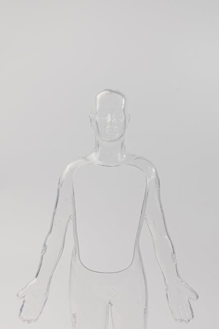
Elevate your anatomy study with interactive platforms and apps designed for visual learners; Tools like Visible Body and Complete Anatomy offer 3D models, allowing you to explore structures from any angle. Interactive dissection simulations and quizzes enhance engagement, while real-time labeling helps identify key anatomical features. Apps like AnatomyTOOL provide virtual labs and case studies, bridging theory with practice. These platforms integrate with photos and videos, offering a comprehensive learning experience. They are ideal for self-paced study, enabling you to master complex anatomical concepts dynamically and effectively, making them indispensable for modern anatomy education.

Regional Anatomy: A Photographic Approach
Explore anatomy through detailed regional photos, from head to limbs. High-quality images reveal structures, aiding visual learners in understanding complex anatomical relationships and clinical applications effectively.
4.1 Head and Neck Anatomy: Key Structures and Photos
Delve into the intricate anatomy of the head and neck through high-resolution photos and detailed dissections. Visualize key structures like the skull, muscles, nerves, and blood vessels. Photos of cadaver specimens provide a realistic view of spatial relationships, while labeled images enhance identification. Clinical correlations, such as CT scans and MRI images, bridge anatomy with real-world applications. This section is ideal for visual learners, offering a comprehensive guide to understanding the complex interconnections of the head and neck region. Use interactive tools to explore 3D models and deepen your grasp of this anatomically rich area.
4.2 Thoracic and Abdominal Anatomy: Visual Guide
Explore the thoracic and abdominal regions through detailed photos and dissections, highlighting key structures such as the rib cage, diaphragm, and abdominal muscles. High-quality images reveal the spatial relationships between organs like the heart, lungs, liver, and intestines. Labeled visuals aid in identifying nerves, blood vessels, and fascia. Clinical correlations, including X-rays and MRIs, provide real-world context. This section emphasizes the importance of visual learning in understanding the complex anatomy of the torso, offering a practical guide for students and professionals alike.
4.3 Pelvic and Limb Anatomy: Detailed Photo Examples
Examine the intricate structures of the pelvic girdle and limbs through high-quality dissection photos. Detailed images reveal the anatomy of the hip joint, knee, and ankle, showcasing muscles, nerves, and blood vessels. Labeled visuals highlight key features like the acetabulum, femur, and tibia. Clinical correlations, such as fractures and joint replacements, provide practical insights. The section also includes photos of hand and foot anatomy, emphasizing their functional complexity. These visual aids are essential for understanding the biomechanics and spatial relationships of the lower body, making them invaluable for both students and professionals studying anatomy.

Systems-Based Anatomy: A Visual Exploration
Explore the human body’s systems through detailed photos and interactive tools, focusing on skeletal, muscular, nervous, and circulatory systems. Visual aids enhance comprehension of complex anatomical relationships.
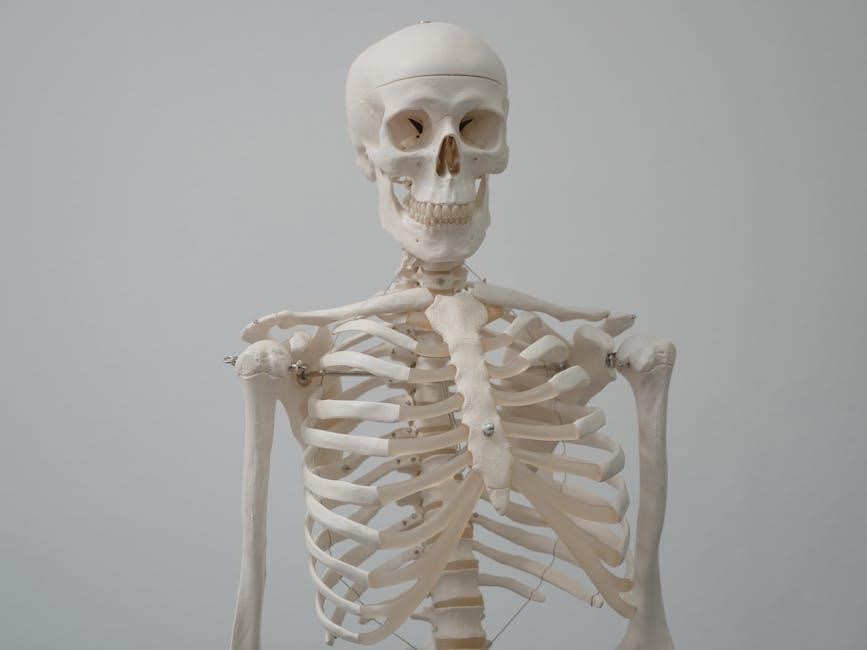
5.1 Skeletal and Muscular System: Photo Atlas
A comprehensive photo atlas of the skeletal and muscular systems, featuring high-resolution images of bones, joints, and muscles. Detailed dissection photos reveal anatomical structures, while 3D models provide interactive exploration. This visual guide highlights the relationship between bones and muscles, showcasing how they work together to enable movement. Included are labeled images of cadaver specimens, illustrating key landmarks and attachments. The atlas also covers clinical correlations, such as common injuries and anatomical variations. With a focus on clarity and precision, this resource is ideal for visual learners aiming to master the intricacies of the skeletal and muscular systems.
5.2 Nervous and Circulatory Systems: Visual Overview
Explore the intricate anatomy of the nervous and circulatory systems through detailed visuals and interactive tools. High-quality images and videos showcase the brain, spinal cord, and peripheral nerves, while diagrams illustrate neural pathways. The circulatory system is brought to life with 3D models of the heart, blood vessels, and blood flow dynamics. This visual guide combines real-time animations and labeled specimens to enhance understanding. Interactive platforms allow users to dissect and examine structures virtually, fostering a deeper connection between anatomy and physiology. Perfect for visual learners, this resource bridges complex concepts with engaging, easy-to-follow content.

Clinical Correlations in Anatomy
Understanding the functional and clinical significance of anatomical structures enhances diagnostic and therapeutic applications. High-quality images and case studies illustrate these connections, aiding in knowledge retention and practical application.
6.1 Surface Anatomy and Its Clinical Relevance
Surface anatomy bridges the gap between gross anatomy and clinical practice, enabling healthcare professionals to identify internal structures through external landmarks. This knowledge is crucial for diagnostic procedures, such as palpation, imaging, and surgery. High-quality photos and 3D models illustrate key surface features, aiding in the localization of organs and anatomical pathways. For instance, understanding the position of the heart valves or liver margins through surface markings enhances physical examination skills. Clinically relevant imaging, like X-rays and MRIs, further correlates surface anatomy with internal structures. This integration is vital for accurate diagnoses, informed treatments, and effective patient care, making it a cornerstone of medical education and practice.
6.2 Diagnostic Imaging: X-rays, MRIs, and CT Scans
Diagnostic imaging is a cornerstone of modern anatomy, providing detailed views of internal structures non-invasively. X-rays are ideal for bone fractures and lung conditions, while MRIs offer superior soft tissue detail, such as muscles and organs. CT scans combine X-ray images to create cross-sectional views, enhancing spatial understanding. These tools are essential for clinical diagnosis, allowing healthcare professionals to identify abnormalities and plan treatments. High-quality images from sources like BlueLink and Visible Body integrate with anatomical studies, aiding in the correlation of surface anatomy with internal structures. This visual integration is vital for both education and patient care, bridging anatomy with real-world applications.

Anatomy of the Human Body: A Photographic Dissection Guide
Explore the human body through detailed dissection photos and interactive tools, offering a step-by-step guide to understanding complex anatomical structures and their relationships.
7.1 step-by-step Dissection Techniques
7.1 Step-by-Step Dissection Techniques
Mastering dissection requires precision and patience. Begin with proper preparation, including gloves, scalpels, and magnifying tools. Start by identifying surface anatomy before making precise incisions. Use forceps to gently expose deeper structures, ensuring minimal damage. Document each step with high-quality photos for later review. Follow a systematic approach, focusing on one region at a time to maintain clarity. Practice on cadaveric specimens or 3D models to refine your skills. These techniques enhance spatial awareness and deepen understanding of anatomical relationships, proving invaluable for visual learners and aspiring health professionals.
7.2 Identifying Key Structures in Cadaver Specimens
Identifying key structures in cadaver specimens requires a systematic approach. Begin by examining the specimen from multiple angles, noting landmarks and relationships. Use forceps to gently expose tissues, ensuring careful handling to avoid damage. Reference high-quality anatomical atlases or digital tools like Visible Body for comparison. Document findings with detailed photos, labeling structures for clarity. Cross-reference with radiographic images to enhance understanding of spatial relationships. This hands-on method fosters deep anatomical knowledge, especially for visual learners. Pay special attention to clinically significant structures, as they often highlight the practical applications of anatomical study.

Learning Aids and Study Tools
Enhance your anatomy study with interactive tools like flashcards, quizzes, and 3D models. Utilize virtual dissection software and photo guides for immersive, visual learning experiences.
8.1 Flashcards and Quizzes for Anatomy Mastery
Flashcards and quizzes are essential tools for reinforcing anatomy knowledge. They promote active recall and spaced repetition, enhancing long-term retention. Apps like Visible Body offer interactive quizzes with 3D models and photo guides, allowing you to test your understanding of anatomical structures. Flashcards often feature high-quality images, labels, and clinical correlations, making them ideal for visual learners. Many platforms also include progress tracking, enabling you to identify and focus on weaker areas. These tools are particularly useful for self-study, providing a structured way to master complex anatomical concepts at your own pace. Utilize them alongside your home lab for comprehensive learning.
8.2 3D Models and Virtual Dissection Tools
3D models and virtual dissection tools revolutionize anatomy study by providing immersive, interactive experiences. These tools allow users to explore anatomical structures in detail, label parts, and engage with simulations. Platforms like Visible Body offer high-quality 3D models that can be rotated, zoomed, and dissected virtually, enhancing spatial understanding. Virtual dissection tools mimic real-life lab experiences, enabling users to identify structures and their relationships; These resources are ideal for visual learners and complement traditional study methods. Accessible online or via apps, they offer flexibility for home lab setups, making complex anatomy concepts more engaging and easier to master.

Advanced Anatomy Topics
Explore advanced anatomy topics through detailed photos and guides. Delve into embryology, histology, and microscopic structures, enhancing your understanding of complex anatomical systems and their clinical correlations.
9.1 Embryology and Developmental Anatomy
Embryology and developmental anatomy explore the formation and growth of the human body from conception to maturity. High-quality images and detailed guides illustrate key developmental stages, including gastrulation, organogenesis, and morphogenesis. This section provides a visual journey through embryonic development, highlighting critical structures and their transformations. Interactive tools and 3D models complement traditional photos, offering deeper insights into complex developmental processes. Understanding these concepts is crucial for appreciating congenital anomalies and their clinical implications. By integrating visual and textual resources, learners can master the dynamic and intricate field of embryology, laying a strong foundation for advanced anatomical studies and practical applications in medicine.
9.2 Histology and Microscopic Anatomy
Histology and microscopic anatomy focus on the study of tissues and cells under a microscope, revealing the intricate structures that form the human body. High-resolution images and interactive platforms provide detailed views of cellular arrangements, aiding in the identification of tissue types and their functions. This section emphasizes the importance of visual learning in understanding microscopic anatomy, offering resources like virtual slides and 3D models. By exploring histological specimens visually, learners can better comprehend the relationship between tissue structure and physiological function, enhancing their ability to apply this knowledge in clinical and research settings effectively.

Case Studies and Practical Applications
Explore real-life anatomical applications through clinical case studies, enhancing your understanding of human structure and function in medical scenarios and diagnostic imaging.
10.1 Clinical Case Studies in Anatomy
Clinical case studies provide real-world applications of anatomical knowledge, connecting theoretical concepts to practical medical scenarios. Through detailed patient histories and diagnostic imaging, learners can observe how anatomical structures relate to clinical conditions. These studies often include X-rays, MRIs, and CT scans, offering a visual understanding of injuries, diseases, and surgical interventions. For instance, case studies on fractures or organ pathologies demonstrate the spatial relationships of tissues and their functional implications. Resources like Grays Clinical Photographic Dissector and Netters Head and Neck Anatomy for Dentistry offer comprehensive visual aids to enhance this learning process. Such tools bridge the gap between classroom anatomy and clinical practice, preparing students for professional environments.
10.2 Anatomy in Medical Specialties: Surgery and Emergency Medicine
Anatomical knowledge is crucial in surgery and emergency medicine, where precise understanding of structures is vital for procedures and diagnoses. Surgical specialties rely on detailed dissection guides and imaging to plan interventions. Emergency medicine uses anatomy to quickly identify injuries or conditions. Resources like Grays Clinical Photographic Dissector and Netters Head and Neck Anatomy for Dentistry provide surgeons with visual references. These tools emphasize spatial relationships and clinical relevance, preparing professionals for real-world scenarios. By integrating anatomy with diagnostic imaging, these resources bridge theory and practice, ensuring accurate and effective patient care in high-stakes medical environments.
Future Trends in Anatomy Education
Explore how AI, machine learning, and virtual reality are revolutionizing anatomy education. These technologies offer immersive 3D exploration and personalized learning, enhancing engagement and accessibility for students globally.
11.1 Virtual Reality and Augmented Reality in Anatomy
Virtual and augmented reality are transforming anatomy education by offering immersive, interactive 3D experiences. These technologies allow students to explore complex anatomical structures in detail, enhancing spatial awareness and retention. With VR, learners can dissect virtual cadavers and visualize systems in real-time, while AR overlays digital models onto physical specimens. These tools enable personalized learning, making anatomy more accessible and engaging. By integrating high-quality visuals and interactive simulations, VR and AR provide a cutting-edge approach to understanding human anatomy, particularly benefiting visual learners in home lab settings. This innovative shift is revolutionizing how anatomy is studied, making it more dynamic and effective than ever before.
11.2 AI and Machine Learning in Anatomy Study
Artificial intelligence and machine learning are revolutionizing anatomy education by enabling personalized learning experiences and enhancing visual understanding. AI-powered tools, such as interactive platforms and virtual dissection software, use machine learning algorithms to adapt to individual learning styles, offering tailored exercises and assessments. These technologies also facilitate advanced image analysis, enabling students to explore anatomical structures in unprecedented detail. By integrating AI-driven simulations and real-time feedback, learners can improve their mastery of complex anatomical concepts. This innovative approach not only enhances retention but also prepares students for future advancements in medical and scientific research, making anatomy study more accessible and effective than ever before.